Jaw cysts are slow-growing, mostly fluid-filled sacs within the bone; they can grow for a long time without causing symptoms and gradually damage the surrounding bone, the teeth and neighboring structures. At our Dental Prime clinic, cysts are assessed with imaging methods; treatment is planned by our surgical team according to the cyst's type and size.
What are jaw cysts, and why do they form?
Most of the cysts seen in the jaws originate from the teeth. The most common type is the radicular cyst, which develops from chronic infection at the root tip; untreated deep decay and teeth that have lost their vitality set the stage for it.
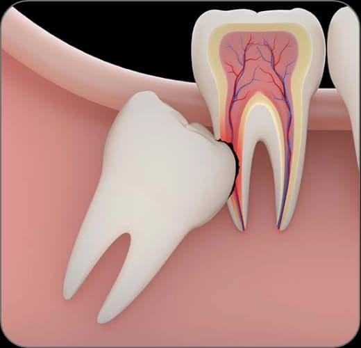
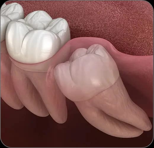
Dentigerous cysts, which develop from the sac around an impacted tooth, are also common; impacted wisdom teeth left without follow-up pose a particular risk in this respect. Because cysts grow slowly, they often go unnoticed for a long time.
Symptoms and diagnosis
A significant share of cysts cause no symptoms at all and are discovered incidentally on X-rays taken during a routine dental check-up. This is one of the situations that shows just how valuable regular check-ups are.
Growing cysts, on the other hand, can announce themselves with swelling of the jaw, a feeling of fullness, shifting or loosening teeth, pain and signs of infection. A panoramic X-ray is the first step in diagnosis; three-dimensional CBCT imaging is used to clarify the cyst's borders and its relationship with the neighboring roots and structures such as the nerve canal or the sinus.
How is cyst surgery performed?
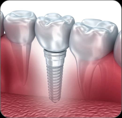
The main goal of treatment is to remove the cyst completely while protecting the surrounding tissues. In most cases the cyst is treated under local anesthesia by removing the sac in one piece (enucleation); depending on the situation, the tooth that caused the cyst is preserved with root canal treatment or extracted.
With very large cysts, a staged approach (marsupialization) that first relieves the cyst's pressure and lets it shrink may be preferred; once the cyst has shrunk, the remaining tissue is removed. In every case the removed tissue is sent for pathological examination; this is a standard and important step for confirming the diagnosis.
Healing and follow-up
After the cyst is removed, the cavity left in the bone fills with new bone over time through the body's own healing capacity; depending on the cyst's size, this process can take several months. In large defects, a bone graft may be used to support healing.
After the operation, X-ray checks are carried out at set intervals; the progress of bone fill and the possibility of recurrence are monitored through these checks. Keeping the follow-up appointments is the final link in the treatment and matters at least as much as the operation itself.
Cysts seen in children
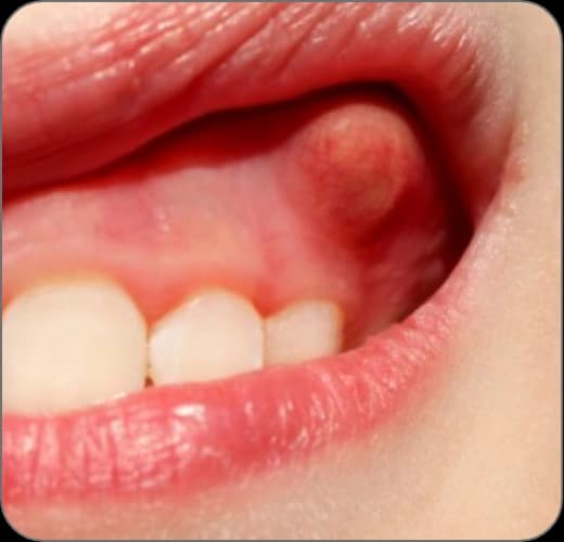
As a pediatric dentist (Assoc. Prof. Dr. Sezin Özer), I occasionally come across a purplish, blister-like swelling over an erupting tooth in my examinations; this is usually a harmless condition we call an eruption cyst, and it generally disappears on its own once the tooth comes through.
Not every swelling in a child therefore requires an operation; in most cases monitoring is enough. Even so, regular check-ups and X-ray follow-up when needed are important for spotting cysts linked to impacted teeth early; in the rare cases that do require surgery, the process is planned gently, according to the child's age and cooperation.
The treatment, step by step
Examination and imaging
The cyst's size, borders and relationship with neighboring structures are determined with a panoramic X-ray and, in cases that require it, a CBCT scan.
Surgical plan and assessment of the tooth
Whether the tooth linked to the cyst can be preserved is decided; root canal treatment or extraction is built into the plan.
Operation under local anesthesia
The cyst is removed while the surrounding tissues are protected; for large cysts a staged shrinking approach may be used.
Pathological examination
The removed tissue is examined in a pathology laboratory to confirm the diagnosis.
Healing and X-ray follow-up
Bone fill and the possibility of recurrence are monitored with check-ups and X-rays at set intervals.