Wisdom teeth are the last teeth to erupt in the mouth; when they cannot find enough room in the jaw, they may remain partially or fully impacted, leading to pain, infection and damage to neighboring teeth. At our Dental Prime clinic, wisdom tooth extraction is planned after a detailed examination and imaging; depending on the tooth's position, a simple or a surgical extraction is performed.
Why do wisdom teeth cause problems?
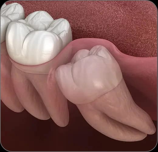
Wisdom teeth usually try to erupt between the ages of 17 and 25. Since the jaw often has too little room left for them, these teeth may erupt at the wrong angle, remain partially impacted or stay completely trapped within the bone.
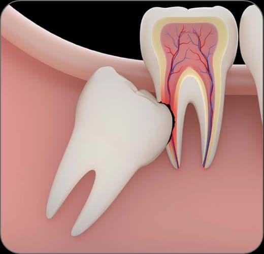
The area under the gum flap covering a partially erupted wisdom tooth cannot be cleaned effectively; bacteria collecting there can trigger recurrent, painful gum infections (pericoronitis). Teeth tilted forward can press on the neighboring molar, causing decay, root resorption and alignment problems.
When is extraction recommended?
Fully erupted wisdom teeth that take part in chewing against an opposing tooth and can be cleaned easily do not need to be extracted. Extraction comes into play when the tooth is causing problems, or is expected to:
- Recurrent gum infections and episodes of pain
- Decay in the wisdom tooth or the neighboring tooth caused by an area that cannot be cleaned
- Pressure on the neighboring tooth, root resorption or crowding
- Signs of a cyst or enlargement in the tissue around the tooth
- When the orthodontic treatment plan requires it
The difference between simple and surgical extraction
A wisdom tooth that has fully erupted in the mouth can usually be removed quickly with a simple extraction after local anesthesia. With partially impacted or impacted teeth, a small incision in the gum, removal of the thin bone over the tooth and sectioning of the tooth may be necessary; this is called a surgical extraction.
Lower wisdom teeth can lie close to the nerve canal that gives sensation to the lip. In cases where this proximity is seen, a three-dimensional CBCT scan maps the course of the nerve and the operation is planned around this information, so that the likelihood of complications such as altered sensation is kept as low as possible.
Healing and care after the extraction
The key to healing after an extraction is protecting the blood clot that forms in the socket. Losing the clot early can lead to a painful condition called dry socket (alveolitis), which is why following a few simple rules in the first days is important.
- Do not spit, rinse or use a straw for the first 24 hours
- Apply intermittent cold compresses externally for the first 48 hours
- Stay away from hot, hard and grainy foods for a few days; prefer lukewarm, soft foods
- Avoid smoking and alcohol; smoking directly impairs wound healing
- Take the recommended medication at the prescribed doses and times
The treatment, step by step
Examination and panoramic X-ray
The tooth's position, eruption angle and relationship with the neighboring teeth are assessed through a clinical examination and a panoramic X-ray.
Three-dimensional imaging when needed
For teeth close to the nerve canal or the sinus, a CBCT scan maps the anatomical structures with millimeter precision.
Anesthesia and extraction
The area is numbed with local anesthesia; a simple or surgical extraction is performed depending on the tooth's position.
Recovery guidance
You receive detailed advice on protecting the clot, on diet and on medication use.
Check-up appointment
After surgical extractions, the stitches are usually removed within a week and healing is checked.