Impacted teeth are teeth that remain within the jawbone or the gum and cannot complete their eruption; over time they can set the stage for pain, infection, damage to neighboring teeth and cyst formation. At our Dental Prime clinic, impacted tooth extraction is planned with detailed imaging and performed comfortably by our surgical team under local anesthesia.
What is an impacted tooth, and why does it stay impacted?
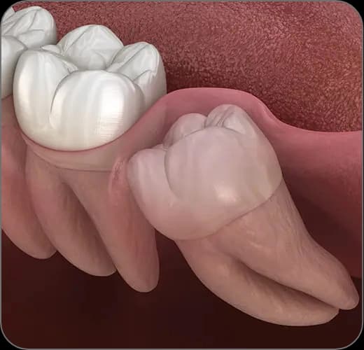
A tooth is considered impacted when it remains partly or completely within the gum or bone even though its time to erupt has come. Wisdom teeth are the most commonly impacted teeth, followed by the canines.
The most common cause of impaction is a lack of space in the jaw. A tooth positioned at the wrong angle, a primary tooth that does not fall out on time, space loss caused by early primary tooth loss and hereditary factors can also block the tooth's path of eruption.
When should an impacted tooth be extracted?
Not every impacted tooth has to be removed. Deeply positioned teeth that cause no symptoms and do not harm the surrounding tissues can be monitored with regular X-ray check-ups. The decision to extract is made by weighing the tooth's position, the patient's age, the symptoms and the relationship with the surrounding tissues together.
Extraction of an impacted tooth is generally recommended in the following situations:
- Recurrent episodes of infection and pain around the tooth (pericoronitis)
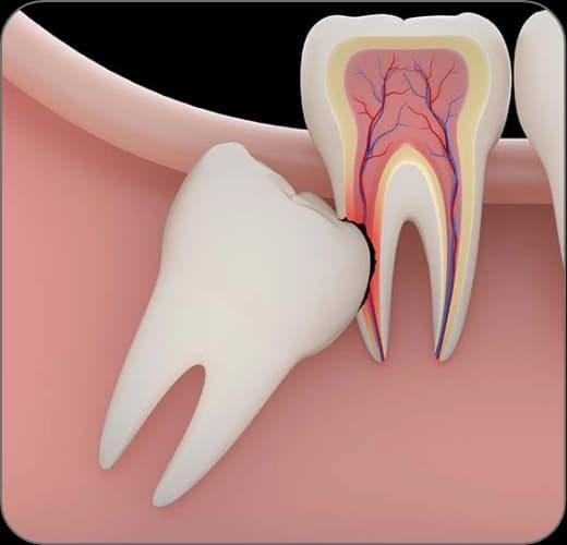
- Decay, root resorption or crowding-related damage in the neighboring tooth
- Enlargement of the sac around the tooth (the follicle) or cyst formation
- When the orthodontic treatment plan requires it
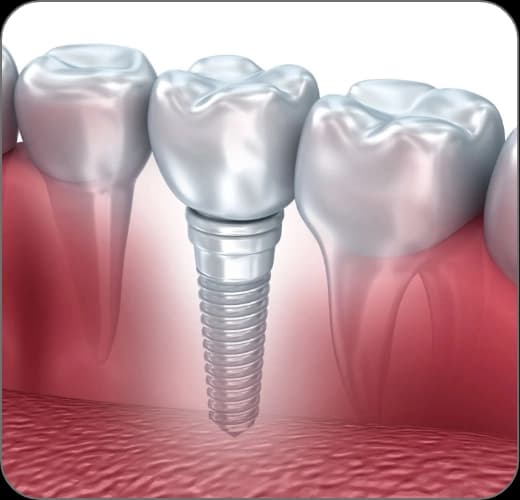
- When the tooth stands in the way of denture or implant planning
How is the operation planned and performed?
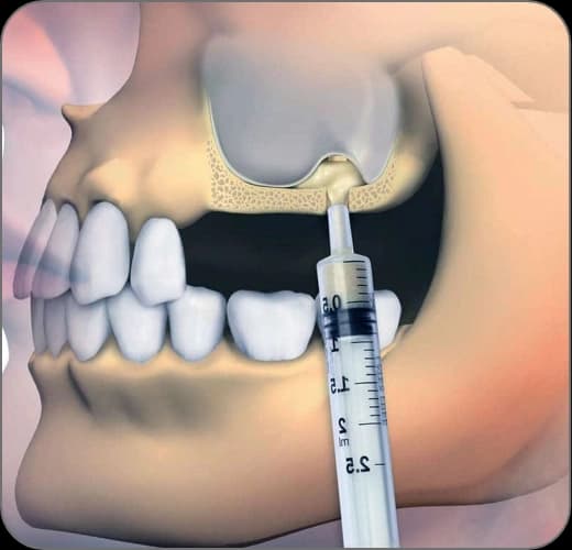
Planning starts with a panoramic X-ray; in cases where the tooth lies close to important anatomical structures such as the nerve canal or the sinus cavity, a three-dimensional CBCT scan allows a millimeter-precise assessment. This way the operation is planned safely, with the neighboring structures protected.
On the day of the operation the area is fully numbed with local anesthesia; the aim is for no pain to be felt during the procedure. A small incision is made in the gum, the thin layer of bone over the tooth is removed if necessary, and the tooth is taken out — sectioned when needed — with respect for the surrounding tissues. After the area is cleaned, stitches are placed to complete the procedure; for a single tooth the operation usually takes around half an hour.
Monitoring impacted teeth in children and adolescents
As a pediatric dentist (Assoc. Prof. Dr. Sezin Özer), I routinely follow the eruption paths of the teeth in my examinations, because the ground for impaction is often laid in childhood. Primary teeth that do not fall out on time, early primary tooth loss and a lack of space can block the eruption path of the permanent teeth.
The tendency of the canines in particular to remain impacted can be spotted early on panoramic films taken during the mixed dentition period. Thanks to early diagnosis, protective steps such as space maintainers, primary tooth extraction or orthodontic guidance can often help the tooth erupt along its own path and reduce the need for surgery later on.
The treatment, step by step
Examination and imaging
After the clinical examination, the tooth's position and its relationship with neighboring structures are assessed with a panoramic X-ray and, in cases that require it, three-dimensional CBCT imaging.
Surgical planning and briefing
The scope of the operation is defined; the procedure, the healing period and the points to watch are explained in detail.
Operation under local anesthesia
The area is fully numbed; the tooth is removed with a tissue-friendly surgical technique and the site is stitched.
Recovery guidance
Advice is given on cold compresses, diet and medication; soft, lukewarm foods are recommended for the first days.
Check-up and suture removal
Healing is assessed at a check-up appointment, usually within a week, and the stitches are removed.