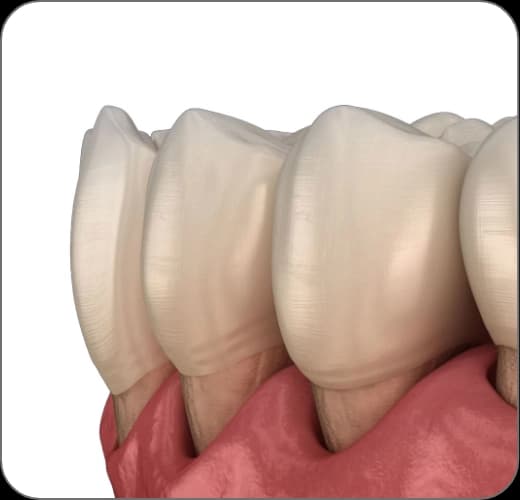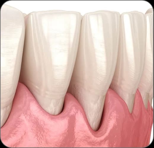
Gum recession — the gradual pulling away of the gum from the root surface — is a common problem that makes teeth look longer, exposes sensitive root surfaces and raises the risk of decay. It is most often the result of advanced gum disease (periodontitis) and, because it progresses silently, tissue may already be lost by the time it is noticed. At our clinic the cause of the recession is identified by our periodontology specialist; treatment follows a plan that extends from controlling the disease to gum grafting where needed.
What causes gum recession?
The most frequent cause is untreated gum inflammation that keeps advancing. When bacterial plaque spreads below the gumline, the body's inflammatory response begins to break down the fibers and bone that support the tooth; as these supporting tissues are lost, the gum pulls away from the root surface and the recession becomes visible.
Beyond disease, personal factors such as aggressive or incorrect brushing, tooth grinding and clenching, poorly fitting restorations and a naturally thin gum type — as well as hormonal phases such as pregnancy and puberty — can all affect the gum tissue. Identifying the underlying cause is the essential first step toward the right treatment plan.
Periodontitis: the disease behind the recession
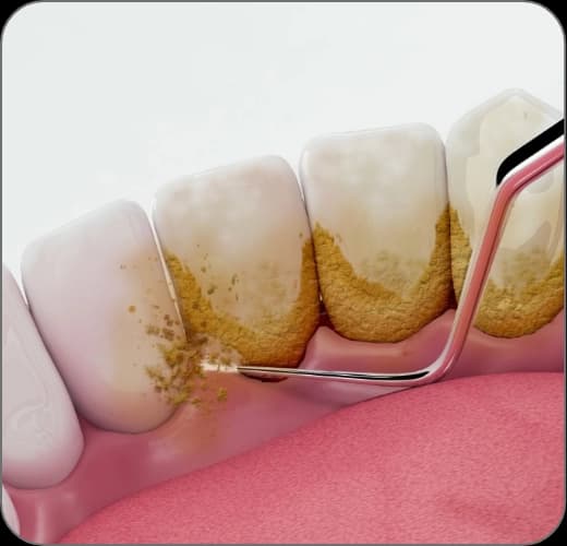
In periodontitis, the most tangible result of tissue destruction is the formation of periodontal pockets. The natural groove around the tooth, normally 1-3 mm deep, deepens into a pathological space as the supporting tissues break down. These deep pockets create sheltered zones that neither toothbrush nor floss can reach; the deeper the pocket, the more bacteria accumulate — and the more bacteria, the faster the bone destruction advances.
The recession cannot be halted without breaking this vicious circle. That is why the first goal of treatment is to bring the disease itself under control: scaling and root planing remove the source of the inflammation, and the healing response is then monitored at regular intervals.
Symptoms: what should you look out for?
Because gum recession usually advances without pain, its signs are easy to miss. If you notice one or more of the findings below, it is advisable to have an evaluation without delay; an early diagnosis gives the chance to intervene before more tissue is lost.
- Teeth that appear longer than they used to
- Increasing root sensitivity to hot and cold
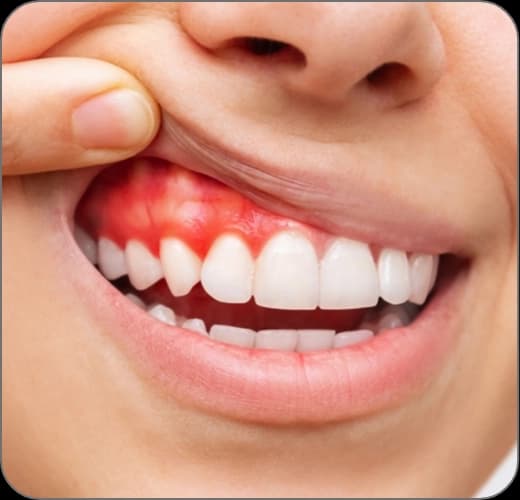
- Thinning, redness or bleeding along the gumline
- New gaps opening up between the teeth
- Loosening or shifting teeth in later stages
Soft tissue grafts: replacing the lost gum
Where the recession has progressed and root surfaces lie exposed, soft tissue grafting (a gum graft) can be used to rebuild the lost tissue. The aim of these procedures is to carry a piece of healthy tissue to the recession site, covering the root surface and reinforcing the gum.
The most commonly used technique is the connective tissue graft, in which a thin piece of connective tissue taken from beneath the surface of the palate is transferred to the recession site. A free gingival graft can be used to increase the amount of protective (keratinized) gum, and where harvesting tissue from the palate is best avoided, processed donor tissue from a tissue bank (an allograft) may be preferred. The right technique is chosen by assessing the type of recession and the quality of the tissues.
When bone has been lost: regenerative options
In cases where periodontitis has caused bone loss, regenerative techniques aimed at regaining the lost supporting tissues may come into consideration when conditions are suitable. In bone grafting, graft materials that encourage new bone formation are placed where the bone has been lost; in guided tissue regeneration, the graft is covered with a special barrier membrane that gives bone cells the time and space to rebuild.
These advanced techniques are not suitable for every case; they give more predictable results particularly in vertical, crater-shaped bone defects. Suitability is determined by evaluating the X-ray findings and the clinical examination together.
The treatment, step by step
Evaluation and cause analysis
Pocket depths are measured and the bone level is examined on X-rays; it is determined whether the recession stems from disease or from mechanical factors.
Controlling the disease
Inflammation is brought under control with scaling and root planing; brushing habits are corrected.
Graft planning
Depending on the type of tissue loss, a connective tissue graft, a free gingival graft or donor tissue options are considered.
The surgical procedure
The procedure is performed under local anesthesia with comfort in mind; the graft is secured over the recession site with fine sutures.
Healing and follow-up
Healing is monitored at regular check-ups; a personalized care program is set up to prevent recurrence.